Vial and blister inspection: every reject needs an image
In medical packaging, "we removed the defective product" isn't enough. You need to show what you removed, why, and when. This post is about how image records change the quality team's work.
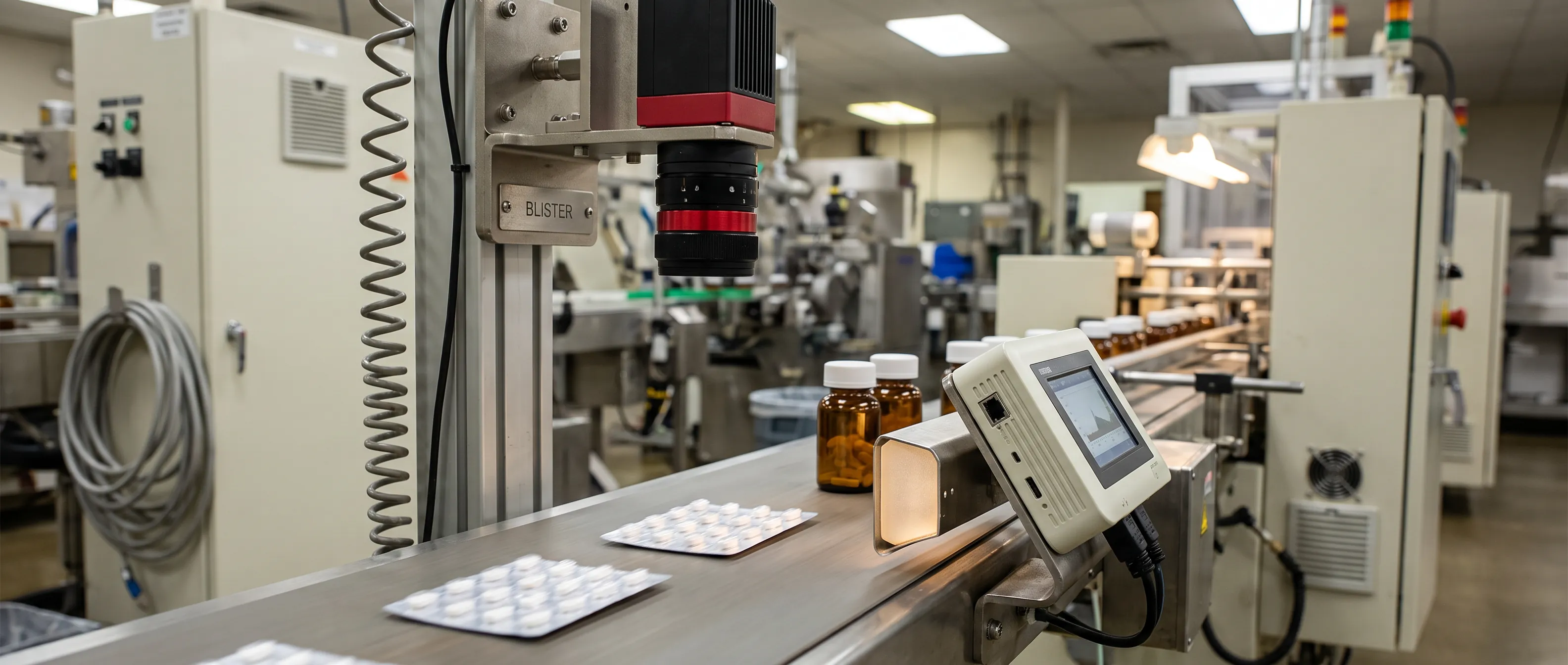
In medical packaging, "we removed the defective product" isn't enough. You need to show what you removed, why, and when. This post is about how image records change the quality team's work.
Quality teams running vial inspection and blister checks hear one question often: "How do you know?" Sometimes it comes from a customer auditor, sometimes from a regulatory inspector, and sometimes from your own QA specialist running a deviation investigation. In medical and pharmaceutical packaging, showing that the product is right matters as much as the product being right.
So this post steps back from "does visual inspection catch the defect?" and asks something else: how does it record what it catches, and what does that record give the quality team?
The difference between a counter and an image
A counter tells you how many products were rejected; an image record shows what was rejected, why, and when. Investigations, internal audits, and customer meetings in medical packaging depend on that second kind of information. And because rejected product is often destroyed, the image is frequently the only evidence left.
On many lines, reject data is just a counter: "312 rejects" at the end of the shift. It doesn't answer your questions. How many of those 312 cards had a missing tablet, and how many a broken one? Were the rejects spread across the shift, or did they cluster after 2 p.m.? Were they all real defects, or were some false alarms?
An image record changes that. Each reject is stored with the original image, the anomaly heatmap marking the deviation, a numeric confidence score, and a time stamp. Behind the number 312 stand 312 pieces of evidence you can look at.
| Question | Counter only | Image record |
|---|---|---|
| How many products were rejected? | Yes | Yes |
| Which defect type? | No | Yes, with the defect label |
| Which pocket of the card? | No | Yes, visible in the image |
| When did rejects cluster? | Only the shift total | By time stamp, minute by minute |
| Real defect or false alarm? | Unknown once the product is destroyed | Judged by looking at the image |
| What do you show an auditor? | A number | Image records and periodic reports |
Blister inspection: which pocket, and what's missing?
The core of blister inspection is judging each pocket, not the card as a whole. Is there a pill in every pocket, is the count right, is it intact? Each is checked, and the image shows which pocket the defect sits in. That location is the shortest route to the root cause.
A pocket with a good tablet is marked OK; a pocket with a missing or broken tablet is marked NG. The screen shows how many good and how many defective pockets that card has. ISEE Vision's AI visual inspection system for pharmaceutical packaging inspection does this across the whole card, on every card. It doesn't sample; it inspects 100%.
The valuable part is the location. Instead of "card rejected," you keep "tablet missing in the second pocket of the third row." If the gap recurs in the same pocket position, it points to a specific feeder channel.
The blister isn't only about tablets. The same station can check gelatin capsules, package damage, bulging, trapped air, cap condition, and folding, gluing, and sealing errors. Serial number, batch code, and expiry date can also be verified, so the record shows the right product left with the right code.
Vial inspection: foreign particles and fill level
Visual inspection of a liquid-filled vial or bottle asks two questions: is there something inside that shouldn't be, and is it filled correctly? A vision system can record foreign matter by type and check fill against a reference. That record supports regulatory compliance; it is not compliance itself.
Inspecting liquid-filled containers is a specialist field, and regulatory expectations reflect that. In Europe, EU GMP Annex 1 (2022) expects filled containers of parenteral products to be inspected individually (§8.30) and sets principles for how that inspection is designed and monitored. In the United States, USP <790> covers visible particulates in injections, and USP <1790> gives guidance on designing the visual inspection of injections.
These texts expect inspection results to be tracked and reviewed, not just produced: which defect types appear, how often, and how the trend moves.
Particle, fiber, glass: why the distinction matters
In our sample work, objects in the liquid were marked one by one and labeled by type: particle, fiber, glass. That distinction matters in an investigation because each has a different source. A fiber usually comes from the environment or a filter. Glass comes from the container itself or from contact during filling. Trapped air is also checked separately; assessing a bubble apart from a real particle helps cut unnecessary rejects.
Fill level and fill ratio
For products such as syrup bottles, the fill ratio is checked from the same image. The system takes a correctly filled bottle as the reference and evaluates the liquid line in each bottle against it and your defined tolerance; a bottle outside the tolerance is marked NG. Where regulation or your own validation requires a certified quantity measurement, keep your existing weighing or level equipment. The visual check complements that record; it doesn't replace it.
A visual inspection system doesn't by itself make you compliant with GMP, Annex 1, or USP expectations. Compliance depends on your processes, validation, and documentation; the system contributes an orderly, image-based record.
What does an image record give the quality team?
An image record gives the quality team four concrete things. It moves deviation investigations from guesswork to evidence, makes false alarms visible, points manual inspection where it is most useful, and documents decisions while leaving them with the quality team. In each case, questions get answered from images, not memory.
Deviation investigations stop being guesswork
When a lot's reject rate climbs, an investigation starts. Without images, the team relies on memory and a few retained samples. With images, you look at the time-stamped rejects for that production window, sort them by defect type, and see when the first deviation began. Daily, weekly, and monthly reports, broken down by defect type, product code, and shift, come from the same records.
False alarms become visible too
In pharma a false reject is costly: good product gets destroyed. Image records let you review false alarms. If the quality team can look at a group of rejects and say "these aren't defects, that's a light reflection," it has hard data for tuning the per-product sensitivity and acceptance threshold.
The limits of human inspection are acknowledged
Manual inspection is still common and valuable, but it has limits. At Sandia National Laboratories, 82 inspectors examined 140 precision-manufactured nuclear-weapons parts for eight defect types and correctly rejected 85% of the defective items (See, Human Factors, 2015). The author notes this is "not vastly superior to the industry average of 80%," so roughly one defect in five gets through typical visual inspection. The high hit rate in this study also came with 35% of good parts rejected, a scrap rate the author calls not typically observed. Even a careful, experienced team can miss defects, and tightening the threshold to catch more risks scrapping good product.
Alongside automated inspection, manual inspection can be pointed where it adds most: a second look at the suspect products the system flags.
The decision stays with the quality team
The system flags a deviation; whether the product is scrapped, re-inspected, or accepted is the quality team's call. The image record supports that decision but doesn't replace it.
Practical points when setting up vial inspection
Results depend on lighting, product teaching, and line response as much as on the algorithm. These come up in our first meeting about vial inspection or blister checks.
Lighting matters as much as software. Aluminum foil, PVC blisters, and glass bottles reflect light very differently, and making a particle in liquid visible may need backlighting or different angles. That is why the station is a light-isolated enclosure, with camera count, lens, lighting angle, and working distance set for each line. Precision goes down to 6 µm, depending on the optical configuration.
No defect catalog needed. ISEE Vision Anomaly Detection learns the good product and flags what deviates, so a deviation nobody has seen before can still be caught. We explain the approach in our post on anomaly detection and its industrial applications.
Format changes are quick. A new blister format or bottle type is taught on site with good samples and saved as a profile under its product code. When you return to it, the operator selects it on the panel; no mechanical adjustment or reinstallation is needed.
You choose how the line responds. Alert only, alert plus line stop through relay or PLC, or a signal to a reject mechanism. Alerts can go to a screen, a tower light, or a siren, and an email can go to the quality lead.
Data stays on site. Processing runs on the station itself, with no cloud dependency, so images don't have to leave the plant. For a data-sensitive industry that is not a small detail; see our post comparing edge AI and cloud systems.
Frequently asked questions
Does a visual inspection system make us GMP compliant?
No, not on its own. GMP and Annex 1 compliance depend on your processes, validation, and documentation as a whole. Time-stamped image records support that documentation and give you evidence you can show in an audit.
What does blister inspection check?
Pill presence and count in each pocket, broken or missing tablets, gelatin capsule condition, package damage, bulging, and cap condition. Serial number, batch code, and expiry date can be verified at the same station.
What line speeds does it handle?
The medical and pharmaceutical station is built for line speeds up to 250 m/min. Real capacity depends on card size, pocket count, and the regions inspected, so we assess it on your line before a pilot.
Can it see a chip or crack in the tablet itself?
Yes. Embossing and the score line are learned as part of the good product, so only a broken edge or an unexpected line is flagged. Our guide to tablet defects covers each defect type.
Key takeaways
A good inspection system in medical packaging can account for everything it removed. With an image behind every reject, the quality team speaks with evidence.
- In blister inspection, the value lies in which pocket and when, not only in the reject.
- In vial inspection, foreign matter is recorded by type and fill is checked against reference and tolerance.
- Image records support GMP documentation but don't deliver compliance on their own.
- Scrap, re-inspect, or accept is always the quality team's decision.
You can see other industries on the automated visual inspection page. If you want to see this on your own blister card or bottle, bring a few good and defective samples and we'll run a free trial on them. Write to us here.
Contact: info@isee-vision.com · +90 216 455 95 44